Monday 29 September 2014

Deep Brain Stimulation Surgery in India, at Low Cost Deep Brain

Deep Brain Stimulation Surgery uses an implanted, battery-operated medical device called a neurostimulator—similar to a heart pacemaker and approximately the size of a stopwatch—to deliver electrical stimulation to targeted areas in the brain that control movement, blocking the abnormal nerve signals that cause tremor and Parkinson’s disease symptoms. The electrode is connected via a wire running beneath the skin to a stimulator and battery pack in the patient's chest. It is reversible—just turns off the current—and allows for precise calibrated symptom control. Before the procedure of Deep Brain Stimulation Surgery, a neurosurgeon uses magnetic resonance imaging (MRI) or computed tomography (CT) scanning to identify and locate the exact target within the brain where electrical nerve signals generate the PD symptoms. 
Deep Brain stimulation may be used in addition to therapy with levodopa or other drugs when drugs alone do not control symptoms adequately. This technique of Deep Brain Stimulation Surgery is the preferred surgical method of treating most cases of advanced Parkinson's disease. It does not destroy brain tissue and has fewer risks than older, more destructive surgical methods, such as pallidotomy and thalamotom
Deep brain stimulation (DBS) is a treatment where a part of your brain is stimulated, to stop you having symptoms of a particular medical condition. It’s called an interventional procedure. An ‘interventional procedure’ includes tests, treatments or surgery which involve making a cut to the skin. Surgery is needed to fit the DBS system.
There have been several studies where people with difficult-to-control epilepsy have had fewer seizures after having DBS surgery. 
  • DBS will only be considered for people who can’t have their seizures controlled by epilepsy medicines or other types of surgery
  • There’s not much good evidence about how well DBS works
  • After two years, more than half the people who had DBS had fewer seizures than before the surgery
  • If you are being considered for DBS, a team of specialist doctors will work together, to make sure you and your epilepsy are suitable for the surgery
  • If you are offered DBS, you should be told that the benefits are uncertain, and the surgery has risks
  • Your doctor should discuss the risks with you, and give you written information before you decide whether to go ahead with DBS surgery
  • Risks include bleeding in the brain, infection, depression and memory problems
What surgery for DBS involves
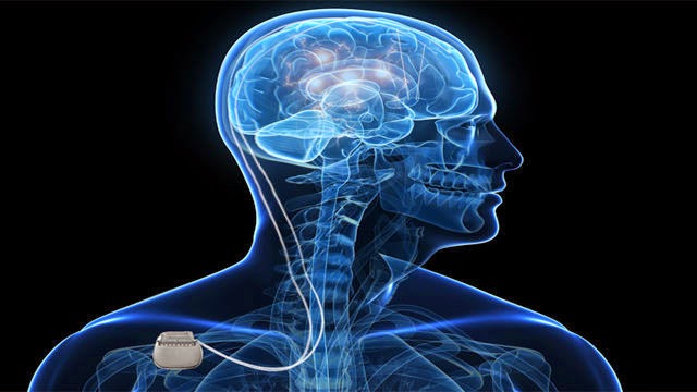
The surgery involves having a DBS system fitted. The DBS system has three parts.
 A lead – this is a thin, insulated wire. It is put through a small opening in your skull, to reach the part of your brain where the epileptic activity happens. Electrical activity is happening in our brain all the time. A seizure happens when there is a sudden burst of intense electrical activity. This is often referred to as epileptic activity.
• An extension – this is an insulated wire that is passed under the skin of your head, neck, and shoulder. It connects the lead to the neurostimulator.
• A neurostimulator – this is a small device, similar to a heart pacemaker. It is usually placed under the skin near your collarbone, lower in your chest, or under the skin of your stomach.
Before the surgery, a brain surgeon will give you a magnetic resonance imaging (MRI) or computed tomography (CT) scan. This is to see the exact part of your brain where the epileptic activity happens.
At the start of the surgery, you will take some drugs to make you relaxed and sleepy, but you might stay awake. You might have a frame attached to your face. The frame will be taken away when the surgery is finished.
After the surgery, the surgeon may give you another CT or MRI scan, to make sure the DBS is in the right place.
What DBS does
Once the DBS is in place, electrical impulses go from the neurostimulator, along the extension wire and lead, and into your brain. These stimulate the part of your brain where there is epileptic activity, to stop your seizures happening. The surgeon will use a programming unit to turn the neurostimulator on, adjust the stimulation, and monitor activity. You will be given a hand held programmer or a magnet, so that you can switch the stimulator on and off.
Deep Brain Stimulation Surgery in India is being dealt by expert Neurosurgeons who have the expertise and many years of experience in performing complex Neuro surgical procedures and have hands on experience with the latest technological devices used to perform the most sophisticated surgeries. The hospitals catering Deep Brain Stimulation Surgery in India have network of hospitals which have dedicated state-of-the-art Neurosurgery operation theaters with semi-robotic microscopes, the neuro-navigation facility, cranial and spinal endoscopy, MRI and CT compatible stereotaxy for functional neurosurgery. It assists the surgeons in attaining precision while conducting complex neurosurgeries.
Why should you choose to get Neurology & Endovascular Neurosurgery in India?
  • Indian doctors are known all over the world for their skill and knowledge and have the experience of studying and working at the best neuron surgery hospitals in the world.
  • Most advanced Technology Infrastructure - Blood Bank with 24 hour apharesis facility, advanced laboratory and microbiology (infection control) support, advanced cardiology, DSA and interventional radiology, portable and colour ultra-sonology, Liver Fibro-scan, 64 slice CT scanner, 3 T MRI, PET-CT and nephrology (including 24 hour dialysis and CVVHD).
  • Neurosurgery Hospitals in India are equipped with the latest and high end technology.
  • Cost of epilepsy surgery in India at best brain surgery hospitals in India is very low as compared to the cost at best hospitals in America or UK with the same level of care and services.

Friday 26 September 2014

Epilepsy should be diagnosed as early as possible to give a child the best chance for treatment success and a normal childhood

Epilepsy in children have many causes. There is an important difference between something that causes seizures, such as a high fever in a young child, and something that causes epilepsy, such as a severe head injury.
Symptoms of epilepsy may be difficult to recognise in children, however: not all seizures involve obvious convulsions. Sometimes symptoms are far more subtle, and these "hidden signs" often appear to fall within the range of normal childhood behaviour. For example, some seizures resemble daydreaming; or a seizure that causes a toddler to stumble and fall may seem quite unremarkable. When these seemingly common behaviours occur unusually often or in patterns, they may mean that a child has epilepsy.
Epilepsy can also be difficult to diagnose in young children because they cannot clearly communicate their subjective experience.
Recognising "hidden signs" in children
Signs that may mean a young child is having seizures include the following:
·         Short spells of blank staring that look like daydreaming.
·         Sudden falls for no apparent reason.
·         Lack of response for brief periods.
·         Dazed behaviour.
·         Unusual sleepiness and irritability when wakened.
·         Repetitive nodding.
·         Rapid blinking.
·         Frequent complaints from the child about things looking, sounding, tasting, smelling or feeling "funny".
·         Clusters of "jackknife" movements in babies sitting down.
·         Clusters of grabbing movements with both arms in babies lying on their backs.
·         Sudden stomach pain followed by confusion and sleepiness.
·         Repeated movements that look out of place or unnatural.
·         Frequent stumbling or unusual clumsiness.
·         Sudden episodes of fear for no apparent reason.

Recognising "hidden signs" in adolescents
Sometimes seizures start in adolescence. These seizures may also be difficult to recognise, and are often misinterpreted as "going through a phase", psychological problems or being under the influence of drugs or alcohol. The following may be signs that a teenager is having seizures:
·         Blank staring, followed by chewing, picking at clothes, mumbling or random movements.
·         Sudden fear, anger or anxiety for no apparent reason.
·         Muscle jerks of the limbs or body, especially in the early morning.
·         Reporting sensory changes: things look, sound, smell or feel strange or different.
·         Memory gaps.
·         Dazed behaviour.
·         Being unable to talk or communicate normally for a short time.
Presence of the signs listed above does not necessarily mean your child has epilepsy, although these symptoms should be brought to a doctor's attention.
The importance of early diagnosis
Recognition of seizures is important for early diagnosis and treatment of epilepsy. Children whose seizures go unnoticed may face problems such as:
·         Learning disabilities: brief blackouts (loss of consciousness) make it hard to follow the teacher's instructions and keep up with lessons in the classroom.
·         Safety risks. Sudden loss of awareness in certain situations, such as swimming, climbing or riding a bicycle, can result in serious injury.
·         Behaviour problems. The child may experience feelings that he or she cannot  communicate, and so becomes frustrated and "acts out".
Social problems. The child and people he or she comes into contact with do not understand the cause of the child's unusual behaviours. The child may withdraw socially or be excluded by other children.

Advanced Epilepsy Treatment Options Available at World Best Hospital inIndia

Till the time a person develops a second seizure, treatment for epilepsy will not begin. This is due to the reason that a person may have one seizure and never develops a second seizure. Medicines are successful in treating epilepsy. These medicines do not cure epilepsy, they prevent recurring of the seizures. These medicines changes the chemical or electrical transmissions in the brain in a particle way that decreases the chance of a seizure.

Vagus Nerve Stimulation (VNS) :
VNS therapy is recommended when medicines are unable to control epilepsy. During the surgical procedure, an electrical device is implanted underneath the skin. The lead of this device is wrapped around one of the nerves on the neck’s left side. This nerve is called as vagus nerve. This device continuously passes an electric dose to the nerve in order to stimulate it. This process reduces the severity and frequency of the seizures. If a person is feeling a warning seizure symptom then extra stimulation is given for preventing recurrence of the seizure.

Ketogenic Diet :  This is also considered as one of the treatment for epilepsy. Ketogenic diet is recommended for children as it involves eating a diet which is low in proteins and carbohydrates and high in fats. The chemical balance of the brain can be altered that also reduces the chance of having seizures. Ketogenic diet is not meant for adults as the diet can result in any other serious medical condition like heart disease, high blood pressure or strokes.

Depending on the particular clinical situation, one of the following surgical procedures is chosen by the Medical team at our affiliate hospital. These operations either aim to resect the disease area of the brain, to disconnect it from other areas so as to relieve the seizures, or to lessen their impact.
  • Anteromedial Temporal Lobectomy
  • Selective Amygdalohippocampectomy
  • Electrocorticography guided Resections
  • Multiple Subpial transections
  • Quadrantic Resections
  • Multilobar Resections
  • Functional Hemispherotomy
  • Vagal Nerve stimulation
  • Radiosurgery (selected cases)


Wednesday 24 September 2014

Why should you choose Indian Hospitals for Brain and Neuro Surgery?

There are many reasons why so many patients from all over the world come to indian hospitals for various types of treatments related to Brain and other Neurological disorders. Following are the most common reasons. 

The term neurosurgery covers both brain and spine surgeries. Basic neurosurgeries in India are performed at more than 2000 hospitals, but the more 
complexneurosurgeries in India are performed at around 200 super specialty top neurosurgery hospitals. Of these, about 20 are top of the line neurosurgery hospitals in India, while the others perform high-end neurosurgeries. These neurosurgery hospitals in India have the latest equipment, highly experienced neurosurgeons and extremely strong support teams as well. The number ofneurosurgeons in India who perform complex neurosurgeries numbers well over 1000.
The large number of neurosurgeries in India enables Indian hospitals to offer low-cost neurosurgery in India. Further, cost of neurosurgery in India is amongst the lowest in the world.
Indian Network Hospitals in India have dedicated Neurology and Neurosurgery Department. The neurosurgeons operate with the most advanced technology systems. They use an image-guided management system for an improved perspective on the progress of complex brain surgeries. It can also help reduce the need for patients to undergo additional surgeries. The neurosurgeons have the expertise in Computer-assisted brain surgery, stereotactic radiosurgery.

Awake Brain Surgery, Deep Brain Stimulation (DBS) and other complex surgical procedures related to brain and nervous system. They have been trained and have worked in some of the leading Brain Surgery Centres across the world. They work with a multi-disciplinary team of doctors that consists of neurologists, neuro-oncologists), neuroradiologists, orthopaedicsurgeons, ENT specialists, endocrinologists, general medicine and rehabilitation, pain medicine, and other areas to provide the most appropriate treatment for your condition.

Intraoperative Magnetic Resonance Imaging (iMRI)

The neurosurgeons at Indin hospitals in India use a high-resolution intraoperative magnetic resonance imaging (iMRI) while performing brain surgeries. This helps them to see the brain tumours while performing surgery thereby greatly reducing the risk of damaging other parts of the brain. Moreover this also helps to confirm the successful removal of the entire tumour. The Intraoperative magnetic resonance imaging (iMRI) is used during the surgery to remove brain tumors and pituitary tumors with greater accuracy and safety. It is also used to place deep brain stimulator systems .

Computer-assisted Brain Surgery

In Computer-assisted Brain Surgery, various imaging technologies, such as Magnetic Resonance Imaging (MRI), Computerized Tomography (CT) and Positron Emission Tomography (PET) are used to create a 3-D model of the brain. The computer system precisely guides the neurosurgeon to the area/s of your brain requiring treatment. This helps the neurosurgeons to plan the safest way to treat your condition with greater degree of accuracy. The computer-assisted surgery helps a lot in performing Awake Brain Surgery. Computer-assisted techniques is also be usedin treating brain tumours and other lesions with precisely focused beams of radiation using stereotactic radiosurgery.

Stereotactic radiosurgery (SRS)

Stereotactic Radiosurgery (SRS) uses precisely focused radiation beams to treat brain tumours and other abnormal growths in the brain. The advantage of modern Stereotactic Radiosurgery is, it delivers high doses of radiation to the tumour with minimal exposure to surrounding healthy tissue. A multidisciplinary team of experts plan the treatment and carefully monitor doses to avoid the risk of radiation overexposure.Unlike traditional radiation therapy, in this treatment, no incision is made. This minimizes the chances of infection or excessive bleeding. A plan has to be developed before undergoing this surgery. A multi-disciplinary team of doctors plan an appropriate treatment for you depending on thetype of tumour, location and size.

Awake Brain Surgery

In an Awake Brain Surgery you're awake during the surgery. Awake Brain Surgery is done by expert brain surgeons to treat some brain conditions such asbrain tumors or epileptic seizures. Awake Brain Surgery allows yourbrain surgeon to work directly with you in order to locate critical functional tissue of the brain. This process is called mapping. Once the brain has been exposed, you will be awakened and the brain is stimulated to determine the function of each area of tissue. You may be awake during surgery to respond to your surgeon. Your responses will help the surgeon to ensure that he is treating the precise area of your brain that needs surgery. Awake Brain Surgery is mainly done if your tumor is near the parts of your brain that control movement or speech. Awake Brain Surgery helps in lowering the risk of damage to functional areas of your brain. In Awake brain surgery the important areas of the brain can be avoided and preserved during surgery.

Minimally Invasive Brain Surgery

Minimally invasive brain surgeries are commonly done at Indian hospitals in India. The hospitals have expert surgeons who specialise in minimally invasive brain surgery techniques. In a minimally invasive brain surgery , the brain surgeons insert two small tubes and use what are called endoscopes. One tube carries a fibre optic camera and light source, the other carries miniaturized surgical instruments. The surgeons operate by watching a TV monitor.
In the traditional way of doing a brain tumour surgery, a more invasive approach was used to remove tumors close to the brainstem. The surgery took much longer time and was performed through an incision behind and below the ear. This took hours of bone drilling to reach the tumor. Since it was an open surgery, the risks were more. Nowadays, with the development of technology and expertise, some of the brain surgeries can be done through nose. The technique is called the endoscopic expanded endonasal brain tumour surgery. 
This technique allows neurosurgeons to pass a camera and specialized surgical instruments to the back of the nasal passage and directly to various regions of the skull base, without scarring and with a shorter recovery. Even though this technique was first described decades ago for pituitary tumor surgery, but recent advancements have helped to use the endonasal approach to safely remove tumors in a variety of other critical skull-base locations. There are very few surgeons in the world who have the expertise to use this technique in brain tumour surgery. At our network hospitals in India, you will find some expert neurosurgeons who have successfully performed many brain tumour surgeries using this technique. Some of the benefits of Endoscopic TransnasalBrain Tumour Surgery are No Brain Retraction, Faster Recovery Time, No Facial Scars, Fewer Complications and shorter Hospital Stays.


Tuesday 16 September 2014

How is Parkinson’s disease treated now and How could stem cells help?

How is Parkinson’s disease treated now?


Current treatments for Parkinson’s include the drug  It is converted into dopamine in the body, so it acts as a stand-in for the lost dopamine-producing neurons. Some other drugs act like dopamine to stimulate the nerve cells. Patients are also treated with occupational therapy, physiotherapy, healthy diet and exercise. Surgery, such as deep brain stimulation with implanted electrodes, is used to treat advanced symptoms.
These treatments relieve the symptoms of Parkinson's disease, but do not slow down or reverse the damage to nerve cells in the brain. Over time, the symptoms often get worse despite treatment. By the time patients are diagnosed with Parkinson’s they have often had the disease for years and have lost most of the critical nerve cells. Tests that detect Parkinson’s earlier may help, but scientists are searching for a way to replace the damaged cells.
How could stem cells help?
Although the underlying cause of Parkinson's disease is unknown, scientists do know which cells and areas of the brain are involved. Researchers are already using stem cells to grow dopamine-producing nerve cells in the lab so that they can study the disease. Because a single, well-defined type of cell is affected, it may also be possible to treat Parkinson’s by replacing the lost nerve cells with healthy new ones.
Replacing lost cells
Doctors and scientists think cell replacement therapy will work because of the results of transplantation studies done in the 1980s. Scientists took cells from the adrenal glands of four Parkinson’s patients and transplanted them into the patients’ brains. The adrenal glands sit on top of the kidneys and contain some cells that release dopamine and similar substances. After the transplants, there was some improvement in the patients’ condition, but it was only minor and did not last long. This was the first time dopamine-producing tissue had been transplanted into the human brain. In later experiments, researchers have transplanted dopamine-producing neurons from human fetuses into animals and human patients, with major improvements in some cases but only modest changes in others However, there were some side effects in one group of patients and in some cases the disease spread to the transplanted fetal cells more than a decade after surgery.
Scientists still hope that introducing young cells into the brain could delay the onset or progress of Parkinson’s disease, but not enough fetal tissue is available to treat the large numbers of Parkinson’s patients, and the use of foetuses also raises ethical questions. Stem cells could offer an alternative source of new cells for Parkinson’s patients:
·         Embryonic stem (ES) cells could be directed to make dopamine-producing neurons, which could be transplanted into patients. Dopamine-producing neurons have been made from both mouse and human embryonic stem cells in the laboratory.

·         Induced pluripotent stem (iPS) cells could be made from a patient’s adult skin cells in the lab, and then used to make dopamine-producing neurons. In 2010 scientists in the USA treated rats with neurons made from human skin cells using iPS techniques. The transplanted neurons improved symptoms of Parkinson's disease in the rats. However, mice and rats require fewer neurons than humans and it is not yet clear whether this approach would work in patients. More studies are also needed to make sure the cells are safe and would not cause tumours in the brain.

Current research
Stem cell treatments for Parkinson's are still in the early stages of development. Some of the most important recent advances include work on methods for making dopamine-producing neurons in the lab; research on how to improve the effectiveness of transplants and avoid side effects; and studies investigating how the disease works and how cells can help with the development of new drugs.


Monday 15 September 2014

What you can expect from Deep brain stimulation : Most Advanced Technology at World Best Hospitals in India

Facts of Deed Brain Stimulation


Deep brain stimulation (DBS) is a surgical procedure used to treat a variety of disabling neurological symptoms—most commonly the debilitating symptoms of Parkinson’s disease (PD), such as tremor,rigidity, stiffness, slowed movement, and walking problems.

·         The procedure is also used to treat essential tremor, a common neurological movement disorder.

·         DBS does not damage healthy brain tissue by destroying nerve cells. Instead the procedure blocks electrical signals from targeted areas in the brain.

·         At present, the procedure is used only for patients whose symptoms cannot be adequately controlled with medications.DBS uses a surgically implanted, battery-operated medical device called aneurostimulator—similar to a heart pacemaker and approximately the size of a stopwatch—to deliver electrical stimulation to targeted areas in the brain that control movement, blocking the abnormal nerve signals that cause tremor and PD symptoms.Before the procedure, a neurosurgeon uses magnetic resonance imaging (MRI) or computed tomography (CT) scanning to identify and locate the exact target within the brain where electrical nerve signals generate the PD symptoms.

·         Some surgeons may use microelectrode recording—which involves a small wire that monitors the activity of nerve cells in the target area—to more specifically identify the precise brain target that will be stimulated.

·         Generally, these targets are the thalamus, subthalamic nucleus, andglobus pallidus.
·         Once the system is in place, electrical impulses are sent from the neurostimulator up along the extension wire and the lead and into the brain. These impulses interfere with and block the electrical signals that cause PD symptoms.

·         Brain surgery. For the brain surgery portion, your care team fits you with a special head frame to keep your head still during the procedure (stereotactic head frame). Then, they use magnetic resonance imaging (MRI) to map your brain and identify the area in your brain where they'll place the electrodes. You'll be given a local anesthetic to numb your scalp before surgery. You won't need an anesthetic in your brain itself because the brain has no pain receptors.
Your surgeon implants a thin wire lead with four contacts (electrodes) at the tips into a specific area of your brain. A wire runs under your skin to a generator (neurostimulator) implanted near your collarbone.
You'll remain awake and alert during the procedure so that your neurologist can talk to you to make sure the right areas of your brain are being stimulated. Your responses help your health care team place the lead in the correct place while minimizing side effects. During surgery, both the neurologist and surgeon carefully monitor your brain to help ensure correct electrode placement.
·         Chest wall surgery. During the second portion of the surgery, the surgeon implants the part of the device that contains the batteries (pulse generator) under the skin in your chest, near your collarbone. This usually occurs a few days after the brain surgery. General anesthesia is used during this procedure. Wires from the brain electrodes are placed under your skin and guided down to the battery-operated pulse generator. The generator is programmed to send continuous electrical pulses to your brain. You control the generator, and you can turn it on or off using a special remote control.
·         After the procedure
After surgery, you'll need to take antibiotics to lower the risk of infection. A few weeks after surgery, the pulse generator in your chest is activated in your doctor's office. The doctor can be easily program your pulse generator from outside your body using a special remote control. The amount of stimulation is customized to your condition.
Stimulation may be constant, 24 hours a day, or your doctor may advise you to turn your pulse generator off at night and back on in the morning, depending on your condition. You can turn stimulation on and off with a special remote control that you'll take home with you. In some cases, your doctor may program the pulse generator to let you make minor adjustments at home.
The battery life of your generator varies with usage and settings. The battery may last between three and five years. When the battery needs to be replaced, your surgeon will replace the generator during an outpatient procedure.
You must remain in close contact with your surgeon and neurologist to make sure that the pulse generator is working correctly.

For more information visit:          http://www.medworldindia.com       
                    
https://www.facebook.com/medworld.india

Please scan and email your medical reports  to us at care@medworldindia.com and we shall get you a Free Medical Opinion from India’s Best Doctors.

Call Us : +91-9811058159
Mail Us : care@medworldindia.com