Friday 27 March 2015

What are the Different kinds of epilepsy in children's

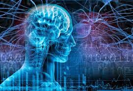
To understand epilepsy, you first have to understand how the brain functions. The brain controls and regulates all voluntary and involuntary responses in the body. It consists of nerve cells that normally communicate with each other through electrical activity.

Epilepsy is defined as two or more seizures that occur without a specific cause. Seizures are altered behavior that occurs when part(s) of the brain receives a burst of abnormal electrical signals that temporarily interrupts normal electrical brain function.

Different kinds of epilepsy are classified according to:

·         Signs and symptoms
·         The child's age when they begin to occur
·         The child’s EEG pattern
·         Neurologic findings on examination
·         Special kinds of imaging (x-ray-type) tests, including magnetic resonance imaging (MRI) and computerized tomography (CT) scans

Seizure Classifications

Focal Seizures

These seizures take place when abnormal electrical brain function occurs in one or more areas of one side of the brain. If the child does not lose consciousness throughout the seizure, the seizure was previously classified as simple. If the child loses consciousness or does not respond appropriately, the seizure was previously classified as complex.

Focal seizures may include one-sided jerking movements of the arms or legs, stiffening, eye deviation to one side or twisting of the body. Sometimes these symptoms follow these other signs:

·         Seeing visions
·         Hearing noises
·         Tasting and smelling things
·         Dizziness
·         A rapid heart rate
·         Dilated pupils
·         Sweating
·         Flushing
·         Stomach fullness
·         Psychic symptoms such as a sense of deja-vu, distortions, illusions and hallucinations

Generalized Seizures

These seizures involve both sides of the brain. As a result, the seizures are less variable than focal seizures.

Typically, they involve brief staring spells; sudden, quick muscle jerks; generalized and rhythmic jerking of the extremities; generalized stiffening episodes; or generalized stiffness followed by rhythmic jerking of the extremities or a sudden loss of muscle tone, resulting in a head drop or sudden fall to the ground.

Causes

Fever (febrile seizures) are caused by fever in children age three months to five years of age, with no other underlying neurologic problems present. Febrile seizures are common and occur in 2-5% of all children. Simple febrile seizures are brief (usually less than five minutes), generalized convulsions that only occur once in the course of an illness.

Metabolic or chemical imbalances in the body may also cause seizures. Conditions that prompt seizures include hypoglycemia (low blood sugar), hypo/hypernatremia (too little or too much sodium in the blood) and hypocalcemia (too little calcium). Meningitis or encephalitis (brain infections) may also induce seizures. Other acute problems that can cause seizures include toxins, trauma and strokes. In children with epilepsy, a common reason for sudden increase in seizures is that the youngsters are not taking their medications as directed.

Trauma at birth or brain abnormalities such as tumors can also be the source of seizures. A lack of adequate oxygen near the time of birth, trauma, infection and stroke can induce a seizure. Sometimes the seizures appear suddenly, although the brain abnormality may have been present for a long time.
Seizures can also develop as a result of a neurodegenerative disease. While neurodegenerative diseases are rare, they can be devastating.

The best tool the doctor has to evaluate the spells is the child's history. This includes knowing what happened immediately before the seizure, the first indication that something was wrong, a complete description of the event, the level of responsiveness of the child, how long the seizure lasted, how it resolved and what the child did after the event. All or some of the following tests may be used:
Blood tests
·         Electroencephalogram (EEG) – A procedure that records the brain's continuous, electrical activity by means of electrodes attached to the scalp.
·         MRI – A diagnostic procedure that uses a combination of large magnets, radiofrequencies and a computer to produce detailed images of organs and structures within the body.
·         CT scan – A diagnostic imaging procedure that uses a combination of x-rays and computer technology to produce cross-sectional images (often called slices) of the body, both horizontally and vertically. A CT scan shows detailed images of any part of the body, including the bones, muscles, fat and organs. CT scans are more detailed than general x-rays.
·         Lumbar puncture (spinal tap) – A special needle is placed into the lower back into the spinal canal. This is the area around (but not into) the spinal cord. The pressure in the spinal canal and brain can then be measured. A small amount of cerebral spinal fluid (CSF) can be removed and sent for testing to determine if there is an infection or other problem. CSF is the fluid that bathes your child's brain and spinal cord.

Medication

Many types of medications may be used to treat seizures and epilepsy. Epilepsy medications are selected based on:
·         The seizure type
·         Child’s age
·         Side effects
·         Consistent use of the medication

Discuss your child's medication side effects with their physician. While your child is taking medications, different tests may be done to monitor the effectiveness of the medication. These tests may include the following:

·         Blood work: Frequent blood draws testing may be required to check the level of the medication in the body. Based on this level, the physician may increase or decrease the dose of the medication to achieve the desired level. This level is called the "therapeutic level," and is where the medication works most efficiently. Blood work may also be done to monitor the effects of medications on body organs.
·         Urine tests: These tests are performed to see how the child's body is responding to the medication.
·         EEG

You should weigh the risks and benefits of therapy versus the risks of a subsequent seizure before your child begins taking a medicine. Treatment is generally not started after the first seizure in children.

While reports vary, the recurrence risk after the first seizure, if it occurred for no apparent reason, is approximately 40%. The majority of recurrent seizures occur soon after the first event — 50% occur within six months.

The use of only one drug is preferable if possible, as approximately 70% of children become seizure-free on therapy with one medication. Another 15% of children become seizure-free on a combination of several. The final 15% have epilepsy that does not respond to medication.

Other Treatments

·         Ketogenic diet – A strict, high-fat diet useful for generalized seizures that don't respond to medication.
·         Vagal nerve stimulator – A surgically implanted wire around the vagal nerve hooked to a pacemaker device in the chest that is programmed to give intermittent stimulation to the vagal nerve. This device is FDA-approved as adjunctive therapy for partial seizures in children over age 12. However, some researchers believe it’s useful for younger children, as well as in children with intractable generalized seizures.

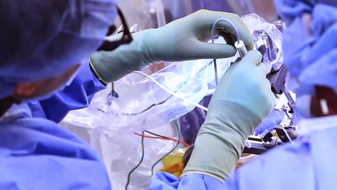
·         Epilepsy surgery – For some patients, particularly those with focal seizures that don't respond to medication or with identifiable lesions on head imaging studies, epilepsy surgery may be the best treatment.
For more information visit:          http://www.medworldindia.com       
                    
https://www.facebook.com/medworld.india
Please scan and email your medical reports  to us at care@medworldindia.com and we shall get you a Free Medical Opinion from India’s Best Doctors.

Call Us : +91-9811058159
Mail Us : care@medworldindia.com

Sunday 22 March 2015

Why should you choose to get Neurology & Endovascular Neurosurgery in India?

Deep Brain Stimulation Surgery uses an implanted, battery-operated medical device called a neurostimulator—similar to a heart pacemaker and approximately the size of a stopwatch—to deliver electrical stimulation to targeted areas in the brain that control movement, blocking the abnormal nerve signals that cause tremor and Parkinson’s disease symptoms. The electrode is connected via a wire running beneath the skin to a stimulator and battery pack in the patient's chest. It is reversible—just turns off the current—and allows for precise calibrated symptom control. Before the procedure of Deep Brain Stimulation Surgery, a neurosurgeon uses magnetic resonance imaging (MRI) or computed tomography (CT) scanning to identify and locate the exact target within the brain where electrical nerve signals generate the PD symptoms. 
Deep Brain stimulation may be used in addition to therapy with levodopa or other drugs when drugs alone do not control symptoms adequately. This technique of Deep Brain Stimulation Surgery is the preferred surgical method of treating most cases of advanced Parkinson's disease. It does not destroy brain tissue and has fewer risks than older, more destructive surgical methods, such as pallidotomy and thalamotom
Deep brain stimulation (DBS) is a treatment where a part of your brain is stimulated, to stop you having symptoms of a particular medical condition. It’s called an interventional procedure. An ‘interventional procedure’ includes tests, treatments or surgery which involve making a cut to the skin. Surgery is needed to fit the DBS system.
There have been several studies where people with difficult-to-control epilepsy have had fewer seizures after having DBS surgery. 
  • DBS will only be considered for people who can’t have their seizures controlled by epilepsy medicines or other types of surgery
  • There’s not much good evidence about how well DBS works
  • After two years, more than half the people who had DBS had fewer seizures than before the surgery
  • If you are being considered for DBS, a team of specialist doctors will work together, to make sure you and your epilepsy are suitable for the surgery
  • If you are offered DBS, you should be told that the benefits are uncertain, and the surgery has risks
  • Your doctor should discuss the risks with you, and give you written information before you decide whether to go ahead with DBS surgery
  • Risks include bleeding in the brain, infection, depression and memory problems
What surgery for DBS involves

The surgery involves having a DBS system fitted. The DBS system has three parts.
 A lead – this is a thin, insulated wire. It is put through a small opening in your skull, to reach the part of your brain where the epileptic activity happens. Electrical activity is happening in our brain all the time. A seizure happens when there is a sudden burst of intense electrical activity. This is often referred to as epileptic activity.
• An extension – this is an insulated wire that is passed under the skin of your head, neck, and shoulder. It connects the lead to the neurostimulator.
• A neurostimulator – this is a small device, similar to a heart pacemaker. It is usually placed under the skin near your collarbone, lower in your chest, or under the skin of your stomach.
Before the surgery, a brain surgeon will give you a magnetic resonance imaging (MRI) or computed tomography (CT) scan. This is to see the exact part of your brain where the epileptic activity happens.
At the start of the surgery, you will take some drugs to make you relaxed and sleepy, but you might stay awake. You might have a frame attached to your face. The frame will be taken away when the surgery is finished.
After the surgery, the surgeon may give you another CT or MRI scan, to make sure the DBS is in the right place.
What DBS does

Once the DBS is in place, electrical impulses go from the neurostimulator, along the extension wire and lead, and into your brain. These stimulate the part of your brain where there is epileptic activity, to stop your seizures happening. The surgeon will use a programming unit to turn the neurostimulator on, adjust the stimulation, and monitor activity. You will be given a hand held programmer or a magnet, so that you can switch the stimulator on and off.
Deep Brain Stimulation Surgery in India is being dealt by expert Neurosurgeons who have the expertise and many years of experience in performing complex Neuro surgical procedures and have hands on experience with the latest technological devices used to perform the most sophisticated surgeries. The hospitals catering Deep Brain Stimulation Surgery in India have network of hospitals which have dedicated state-of-the-art Neurosurgery operation theaters with semi-robotic microscopes, the neuro-navigation facility, cranial and spinal endoscopy, MRI and CT compatible stereotaxy for functional neurosurgery. It assists the surgeons in attaining precision while conducting complex neurosurgeries.
Why should you choose to get Neurology & Endovascular Neurosurgery in India?
  • Indian doctors are known all over the world for their skill and knowledge and have the experience of studying and working at the best neuron surgery hospitals in the world.
  • Most advanced Technology Infrastructure - Blood Bank with 24 hour apharesis facility, advanced laboratory and microbiology (infection control) support, advanced cardiology, DSA and interventional radiology, portable and colour ultra-sonology, Liver Fibro-scan, 64 slice CT scanner, 3 T MRI, PET-CT and nephrology (including 24 hour dialysis and CVVHD).
  • Neurosurgery Hospitals in India are equipped with the latest and high end technology.
  • Cost of epilepsy surgery in India at best brain surgery hospitals in India is very low as compared to the cost at best hospitals in America or UK with the same level of care and services.

Thursday 12 March 2015

Types of Stroke and Treatment in India

Stroke refers to a brain disease caused by either blockage of blood supply or rupture of a blood vessel to a particular part of the brain. This results in reduction of blood and oxygen supply to the affected part of brain resulting in loss of function
A stroke can be categorized into two types –
Hemorrhagic Strokes: This type of stroke occurs when a blood vessel bleeds and burst within the brain. This accumulated blood compresses the nearby tissues of the brain. This could be due to –
  • A blood vessel on the brain surface starts bleeding in between the area of the skull and the brain.
  • A blood vessel bleeding or bursting inside the brain
Hemorrhagic strokes has two main types –
  • Subarachnoid hemorrhage: In this, there is bleeding in between the area of the brain and the thin tissues covering the brain.
  • Intracerebral hemorrhage: This is considered as the most common type of hemorrhagic strokes. This occurs when an artery bursts inside the brain and resulting in the flooding of the nearby blood tissues.
Ischemic Stroke: This occurs when something is causing blockage in an artery which is carrying blood to the brain. The potential causes are –
  • When there is a blockage in the small blood vessels within the brain
  • When a blood clot develops in the main artery to the brain
  • When an air bubble, a blood clot or fat globule develops in a blood vessel which is carried to the brain.
Causes
Some of the causes that can lead to a stroke are as follows:
1.       Ageing
2.      High blood pressure
3.      Diabetes
4.      Cardiovascular diseases
5.      High cholesterol
6.      Obesity
7.      Vitamin B12 deficiency
8.     Excessive alcohol and drug abuse


Symptoms:
Symptoms of a stroke occur suddenly. Symptoms vary depending on the location of the stroke. Each area of the brain is supplied by specific arteries. If an artery supplying the area of the brain that controls the left arm movements is blocked, that arm becomes weak or paralyzed.

The five most common signs and symptoms of stroke are:
  1. Sudden numbness or weakness of the face, arm, or leg - like weakness of half of the body called as hemiplegia.
  2. Sudden confusion or trouble speaking or understanding others called aphasia in medical terms.
  3. Sudden trouble seeing in one or both eyes called as mono or binocular vision disturbance. This could be due a stroke anywhere in the visual pathway.
  4. Sudden dizziness, trouble walking, or loss of balance or coordination, this is commonly due to posterior circulation strokes.
  5. Sudden severe headache with no known cause is due to hemorrhage or sometimes infarction.
Diagnosis:

The diagnosis Is based on the symptoms and a good history given by the patient or an observer. Usually any neurologic symptom occurring suddenly in a neurovascular territory is attributed to a stroke. CT scan done immediately after a stroke is almost always normal in ischemic strokes. MRI with diffusion images is sensitive to diagnose acute infarction.


The treatment for both hemorrhagic and ischemic stroke is different.
The purpose of treating ischemic stroke is to restore the flow of the blood in the brain. Medications are given in the first four and a half hours of the stroke for destroying the clots in the blood vessels. An injection of tissue plasminogen (TPA) can also be given to a patient for improving the probability of a full recovery.
The main purpose of treating a hemorrhagic stroke is to stop bleeding and decrease the pressure on the brain. Instead of blood thinners, clotting drugs can be given. After the area has been healed and the bleeding is controlled, the damaged and leaky blood vessels are then repaired.
The first stroke device approved by FDA is Merci retriever. The aim of this device is to restore the flow of the blood in the neurovasculature by removing thrombus in those patients who have ischemic stroke.
The penumbra system (endovascular thromboaspiration) is the last FDA approved device that has been primarily developed for removing a clot in the case of acute ischemic stroke. In order to eliminate or reduce the clot burden, this system uses dual approaches to clot extraction by using debulking and aspiration of the thrombus. Clot retrieval is then used where a ring device holds the thrombus by capturing it in clasps with a cylinder. This is then withdrawn at the time of flow arrest. This new aspiration device (the penumbra system) has an excellent safety profile and a high rate of ‘target vessel’ recanalization.

For more information visit:          http://www.medworldindia.com      
                    
https://www.facebook.com/medworld.india

Please scan and email your medical reports  to us at care@medworldindia.com and we shall get you a Free Medical Opinion from India’s Best Doctors.

Call Us : +91-9811058159
Mail Us : care@medworldindia.com


Tuesday 10 March 2015

Advanced Epilepsy Treatment Options Available at World Best Hospital in India

How is Epilepsy treated?

The main line of treatment is with antiepileptic drugs, which are effective in controlling seizures in 70%-80% of patients with epilepsy. There are several antiepileptic medications. Since certain medications are much better for some seizures, the choice of the medication should be made by a physician who is familiar with these medications. If possible, the child should be evaluated in a center specializing in epilepsy. If this is not feasible, usually pediatric neurologists have training in epileptic disorders and are a good source for a referral.
When antiepileptic drugs fail to control the seizures, the patients may improve with surgical procedures.
Who is a candidate for epilepsy surgery?

Surgery is indicated in a small group of children.
It usually takes the failure of two or three antiepileptic medications before a child would be considered as a potential candidate for surgery. In general, this happens at least after two or three years of continuous treatment with medications. The failure might be due:
1.   to a resistance to the antiepileptic medications that are available,
2.   to the presence of intolerable side effects to the antiepileptic medication,
3.   or to a combination of both.

Since surgical procedures might be very effective in some children, once it is clear that the child's epileptic disorder is not responding to treatment with antiepileptic medications, surgery should be considered. Young age is not a contraindication for surgery, and there is no benefit in waiting for the child to be older.

In fact, there is considerable evidence that the younger a child is at the time of surgery, the better his/her potential will be for good function after the surgery. There is a certain degree of plasticity in the brain that helps with the recovery of functions that can be damaged at the time of surgery. This plasticity is higher in younger than in older children.
Advanced Epilepsy Treatment Options Available at World Best Hospital in India

Till the time a person develops a second seizure, treatment for epilepsy will not begin. This is due to the reason that a person may have one seizure and never develops a second seizure. Medicines are successful in treating epilepsy. These medicines do not cure epilepsy, they prevent recurring of the seizures. These medicines changes the chemical or electrical transmissions in the brain in a particle way that decreases the chance of a seizure.

Vagus Nerve Stimulation (VNS) :

VNS therapy is recommended when medicines are unable to control epilepsy. During the surgical procedure, an electrical device is implanted underneath the skin. The lead of this device is wrapped around one of the nerves on the neck’s left side. This nerve is called as vagus nerve. This device continuously passes an electric dose to the nerve in order to stimulate it. This process reduces the severity and frequency of the seizures. If a person is feeling a warning seizure symptom then extra stimulation is given for preventing recurrence of the seizure.

Ketogenic Diet :  This is also considered as one of the treatment for epilepsy. Ketogenic diet is recommended for children as it involves eating a diet which is low in proteins and carbohydrates and high in fats. The chemical balance of the brain can be altered that also reduces the chance of having seizures. Ketogenic diet is not meant for adults as the diet can result in any other serious medical condition like heart disease, high blood pressure or strokes.

Advanced Surgical Procedures for Management of Epilepsy

Depending on the particular clinical situation, one of the following surgical procedures is chosen by the Medical team at our affiliate hospital. These operations either aim to resect the disease area of the brain, to disconnect it from other areas so as to relieve the seizures, or to lessen their impact.


  • Anteromedial Temporal Lobectomy
  • Selective Amygdalohippocampectomy
  • Electrocorticography guided Resections
  • Multiple Subpial transections
  • Quadrantic Resections
  • Multilobar Resections
  • Functional Hemispherotomy
  • Vagal Nerve stimulation
  • Radiosurgery (selected cases)


    For more information visit:          http://www.medworldindia.com       
                        
    https://www.facebook.com/medworld.india

    Please scan and email your medical reports  to us at care@medworldindia.com and we shall get you a Free Medical Opinion from India’s Best Doctors.

    Call Us : +91-9811058159
    Mail Us : care@medworldindia.com

Wednesday 4 March 2015

What Are the Symptoms of a Brain Tumor in Adults? : BRAIN TUMOR TREATMENT IN INDIA


Symptoms of brain tumors vary according to the type of tumor and the location. Because different areas of the brain control different functions of the body, where the tumor lies affects the way symptoms are manifested.
Some tumors have no symptoms until they are quite large and then cause a serious, rapid decline in health. Other tumors may have symptoms that develop slowly.
A common initial symptom of a brain tumor is headaches. Often, they don't respond to the usual headache remedies. Keep in mind that most headaches are unrelated to brain tumors.


Other symptoms include:
·         Seizures
·         Changes in speech or hearing
·         Changes in vision
·         Balance problems
·         Problems with walking
·         Numbness or tingling in the arms or legs
·         Problems with memory
·         Personality changes
·         Inability to concentrate
·         Weakness in one part of the body

It's important to keep in mind that these symptoms can be caused by a number of different conditions. Don't assume you have a brain tumor just because you experience some of them. Check with your doctor.
To diagnose a brain tumor, the doctor starts by asking questions about your symptoms and taking a personal and family health history. Then he or she performs a physical exam, including a neurological exam. If there's reason to suspect a brain tumor, the doctor may request one or more of the following tests:
·      Imaging studies such as a CT(CAT) scan or MRI to see detailed images of the brain
·     Angiogram or MRA, which involve the use of dye and X-rays of blood vessels in the brain to look for signs of a tumor or abnormal blood vessels
The doctor may also ask for a biopsy to determine whether or not the tumor is cancer. A tissue sample is removed from the brain either during surgery to remove the tumor or with a needle inserted through a small hole drilled into the skull before treatment is started. The sample is then sent to a lab for testing.

  • Tumors near the surface of the brain may be surgically removed. In many cases, however, it is only possible to remove a portion of a tumor, since taking it all out would cause unacceptable amounts of brain damage. Still, removing even part of it may afford a period of improvement by relieving pressure within the cranium.
  • Tumors deep within the brain may be treated with microsurgery, laser surgery, or radiation therapy.
  • For malignant primary tumors, surgery may be followed with radiation or chemotherapy. Surgery may also be preceded by radiation.
  • Your doctor may prescribe corticosteroids to reduce swelling of brain tissue, anticonvulsant drugs to control seizures, and pain relievers.
Treatment for brain tumors is based on many factors, such as:
  • Your age, overall health, and medical history
  • The type, location, and size of the tumor
  • How likely the tumor is to spread or recur
  • Your tolerance for specific medications, procedures, or therapies
Treatment for these symptoms may include:
  • Antiseizure/Antiepileptic Drugs (AEDs)
  • Steroids
  • Surgery
Often, low-grade tumors (grade I and II), which are not aggressive, are treated with watchful monitoring or surgery alone. Though all tumors are monitored with repeat scans, grade II tumors are watched more closely after surgery and over time to make sure there is no recurrence.
Higher grade tumors (grade III and IV), which are malignant and can grow quickly, are more difficult to remove and require additional treatments beyond surgery, such as radiation, chemotherapy, or a clinical trial if one is available. Microscopic tumor cells can remain after surgery and will eventually grow back. All treatments, therefore, are intended to prolong and improve life for as long as possible.
Additional treatment options for high-grade tumors include:

  • Radiation therapy: X-rays and other forms of radiation can destroy tumor cells or delay tumor growth.
  • Chemotherapy: The use of drugs to kill rapidly dividing cells. It can be taken orally or intravenously.
  • Targeted therapy: The focus on a specific element of a cell, such as molecules or pathways required for cell growth, in order to use them as a target